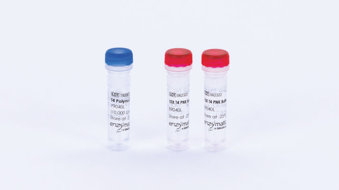
T4 Polynucleotide Kinase (10,000 U)
Cat. No. / ID: Y9040L
Features
- Incorporates labeled phosphates at the 5’ end of nucleic acids
- Catalyzes transfer and exchange of phosphate to the 5′-OH of ssDNA and RNA
- Catalyzes transfer and exchange of phosphate to the 5′-OH of dsDNA
- Removes 3′ phosphoryl groups from DNA and RNA or a nucleotide/nucleoside
Product Details
T4 Polynucleotide Kinase (PNK) is widely used to phosphorylate the 5’ end of both DNA and RNA in labeling and ligation experiments. T4 PNK catalyzes the transfer and exchange of the terminal gamma position phosphate of ATP to the 5′-hydroxyl terminus of double- and single-stranded DNA, RNA and nucleoside 3′-monophosphate molecules.
T4 PNK also catalyzes the hydrolytic removal of 3′-PO4 termini from DNA, RNA and deoxynucleoside 3′-monophosphates. The enzyme is used to remove 3′-phosphoryl groups from nucleotide/nucleoside or DNA and RNA in studies of gene expression and gene function.
In addition to its importance in molecular biology and biochemistry research, T4 PNK exemplifies a family of bifunctional enzymes with 5′-kinase and 3′-phosphatase activities that play key roles in RNA and DNA repair.
The enzyme is supplied in 10 mM Tris-HCl, 50 mM KCl, 0.1 µM ATP, 1 mM DTT, 0.1 mM EDTA, 50% glycerol; pH 7.4 at 25°C.
The enzyme is supplied with a 10X PNK Buffer (B9040) containing 700 mM Tris-HCl, 100 mM MgCl2, 50 mM DTT; pH 7.6 at 25°C.
Performance
| Test | Specification |
| Purity | >99% |
| Specific activity | 133,333 U/mg |
| Single-stranded exonuclease | 2000 U <5.0% released |
| Double-stranded exonuclease | 2000 U <1.0% released |
| Double-stranded endonuclease | 2000 U = No conversion |
| E. coli DNA contamination | 2000 U; <10 copies |
Principle
T4 Polynucleotide Kinase (PNK) catalyzes the transfer and exchange of the terminal gamma position phosphate of ATP to the 5′-hydroxyl terminus of double-and single-stranded DNA, RNA and nucleoside 3′-monophosphate molecules. T4 PNK also exhibits 3′-phosphatase and 2′, 3′-cyclicphosphodiesterase activities.
Procedure
Instructions for using T4 Polynucleotide Kinase are provided in the corresponding kit protocol in the resources below.
Quality Control
Unit activity was measured using a 2-fold serial dilution method. Dilutions of enzyme were made in 1X Reaction Buffer and added to 50 µL reactions containing 10 µM Oligo(dT) single-stranded DNA, 1X PNK Reaction Buffer, and 66 µM ATP and [γ32P]-ATP. Reactions were incubated for 30 minutes at 37°C, plunged on ice, and analyzed using the method of Sambrook and Russell (Molecular Cloning, v3, 2001, pp. A8.25-A8.26).
Protein concentration was determined by OD280 absorbance A 2.0 μL sample of enzyme was analyzed at OD280 using a spectrophotometer standardized using a 2.0 mg/mL BSA sample and blanked with product storage solution.
Physical purity was evaluated by SDS-PAGE of concentrated and diluted enzyme solutions followed by silver-stain detection. Purity was assessed by comparing the aggregate mass of contaminant bands in the concentrated sample to the mass of the protein of interest band in the diluted sample.
Single-stranded exonuclease activity was determined in a 50 μL reaction containing 10,000 cpm of radiolabeled single-stranded DNA and 10 μL of enzyme solution. Incubation for 4 hours at 37°C resulted in less than 5.0% release of TCA-soluble counts.
Double-stranded exonuclease activity was determined in a 50 μL reaction containing 5,000 cpm of a radiolabeled double-stranded DNA substrate and 10 μL of enzyme solution. Incubation for 4 hours at 37°C resulted in less than 1.0% release of TCA-soluble counts.
Double-stranded endonuclease activity was determined in a 50 μL reaction containing 0.5 μg of pBR322 DNA and 10 μL of enzyme solution. Incubation for 4 hours at 37°C resulted in no visually discernible conversion to nicked circular DNA as determined by agarose gel electrophoresis.
E. coli contamination was evaluated using 5 μL replicate samples of enzyme solution denatured and screened in a TaqMan qPCR assay for the presence of contaminating E. coli genomic DNA using oligonucleotide primers corresponding to the 16S rRNA locus.
Applications
This product is available for molecular biology applications such as:
- labeling the 5′ ends of DNA or RNA for use
- as hybridization probes
- in transcript mapping with nucleases
- in sequencing
- phosphorylating oligodeoxynucleotide linkers or other DNA molecules prior to ligation
- end-labeling oligodeoxynucleotide primers for use in sequencing reactions
- dephosphorylating the 3′-ends of RNA in the absence of ATP
- in an exchange reaction to label the 5′ ends of DNA or RNA molecules that have unlabeled 5′ phosphates

