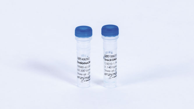
Uracil Cleavage System (10x)
Cat. No. / ID: Y9180L
Features
- Two-enzyme system, Uracil DNA Glycosylase (UDG) and Endonuclease VIII
- UDG catalyzes the excision of the uracil base, creating an abasic site with an intact phosphodiester backbone
- Endonuclease VIII breaks the phosphodiester backbone both 3’ and 5’ to the abasic site, liberating the deoxyribose sugar
Product Details
The Uracil Cleavage System provides two enzymes, which, when added sequentially to a reaction containing a synthetic DNA fragment containing a deoxy-uracil, generate a single nucleotide gap at the location of the uracil residue. The two individual enzyme components, Uracil DNA Glycosylase and Endonuclease VIII, provided at a 10x concentration, are to be added to a reaction containing a uracil-containing polynucleotide sequence. UDG catalyzes the excision of the uracil base, creating an abasic site with an intact phosphodiester backbone (1–2). The lyase activity of Endonuclease VIII breaks the phosphodiester backbone, both 3ʹ and 5ʹ, to the abasic site, liberating the deoxyribose sugar (3–4).
UDG is supplied in 10 mM Tris-HCl, 50 mM NaCl, 1 mM DTT, 0.1 mM EDTA and 50% glycerol; pH 7.5 at 25°C.
Endonuclease VIII is supplied in 10 mM Tris-HCl, 250 mM NaCl, 0.1 mM EDTA and 50% glycerol; pH 8.0 at 25°C.
Performance
| Test | Amount tested | Specification |
| Purity | n/a | >99% |
| Single-stranded exonuclease | 10 μL | <5% released |
| Double-stranded exonuclease | 10 μL | <1% released |
| Double-stranded endonuclease | 10 μL | No conversion |
| E. coli DNA contamination | 5 μL | <10% copies |
Principle
The proteins are produced and purified separately from E. coli strains containing recombinant Endonuclease VIII and Uracil-DNA Glycosylase genes.
One unit of UDG is defined as the amount of enzyme that catalyzes the release of 1.8 nmol of uracil in 30 minutes from double-stranded, tritiated, uracil-containing DNA at 37°C in 1x UDG Reaction Buffer.
One unit of Endonuclease VIII is defined as the amount of enzyme required to cleave 1 pmol of an oligonucleotide duplex containing a single AP site in 1 hour at 37°C.
Procedure
Usage Instructions
The volume can be adjusted to suit the application requirement by maintaining the ratios of components outlined below for a 10 µL reaction.
1. Prepare Uracil-containing DNA (e.g., PCR amplification product). The Uracil Cleavage System enzymes are active in most molecular biology reaction buffers, so there is no need to exchange buffers before assembling the cleavage reaction.
2. Add 0.5 µL UDG (G5010L) to a reaction vessel containing the DNA substrate (final concentration up to 300nM) in 1x reaction buffer.
3. Add 0.5 µL Endonuclease VIII (Y9080L) to the above.
4. Incubate at 37°C for 30 minutes.
5. Following the incubation period, the reaction can be stopped by adding a gel-loading stop solution in preparation for electrophoretic analysis or prepared for transformation. In the case of transformation, the fragment may be ligated into a cloning vector using T4 DNA Ligase (L6030-HC).
Quality Control
Specific activity of UDG was measured using a twofold serial dilution method. The enzyme was diluted in 1x reaction buffer and added to 50 µL reactions containing 1x UDG Reaction Buffer and a PCR product, a 3H-dUTP. Reactions were incubated for 10 minutes at 37°C, placed on ice and analyzed using a TCA-precipitation method.
Specific activity of Endonuclease VIII was measured using a twofold serial dilution method. Dilutions of the enzyme were made in 1x reaction buffer and added to 10 µL reactions containing 2.0 µM of a FAM-labeled, 34-base duplex oligonucleotide, which contained a single uracil. The substrate was pre-treated for 2 minutes with UDG to create an abasic site. Reactions were incubated for 60 minutes at 37°C, placed on ice, denatured with N-N-dimethyl -formamide and analyzed on a 15% TBE-urea acrylamide gel.
Physical purity is evaluated by SDS-PAGE of concentrated and diluted enzyme solutions followed by silver-stain detection. Purity is assessed by comparing the aggregate mass of contaminant bands in the concentrated sample to the band's mass corresponding to the protein of interest in the diluted sample.
Single-stranded exonuclease is determined for each enzyme separately in a 50 µL reaction containing a radiolabeled single-stranded DNA substrate and 10 µL of enzyme solution incubated for 4 hours at 37°C.
Double-stranded exonuclease activity is determined for each enzyme separately in a 50 µL reaction containing a radiolabeled double-stranded DNA substrate and 10 µL of enzyme solution incubated for 4 hours at 37°C.
Double-stranded endonuclease activity v in a 50 µl reaction containing 0.5 µg of plasmid DNA and 10 µL of enzyme solution incubated for 4 hours at 37°C.
E. coli contamination is evaluated using 5 µL replicate samples of enzyme solution that are denatured and screened in a TaqMan qPCR assay for the presence of contaminating E. coli genomic DNA using oligonucleotide primers corresponding to the 16S rRNA locus.
Applications
This product is available for molecular biology applications such as:
- Cloning
References
- Lindhal, T., et al. (1977) J. Biol. Chem. 252:3286.
- Lindhal, T. (1982) Annu. Rev. Biochem. 51:61.
- Melamede, R.J., et al. (1994) Biochemistry 33:1255.
- Jiang, D., et al. (1997) J. Biol. Chem. 272:32230.

